A lot the important things about your mouth are microscopically small. Special instruments are needed to work with them.
The Dental Microscope:
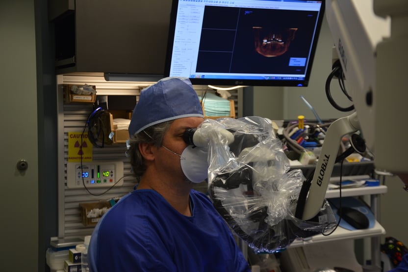
A dental operating microscope (D.O.M.) has been a key tool for endodontic (root canal) dentistry since it became a requirement for residencies to teach specialists to use it in 1998. Over the last 20 years it has been adopted for work in all phases of dentistry including periodontal (gum disease) treatment and restorative dentistry and surgery over the last two decades. These microscopes are practically designed for viewing during actual clinical work with patients.
Advantages of Microscope Dentistry:
The Microscope provides 400 times more visual accuracy than the naked eye and 100 times the visual information than traditional dental loupes. Magnification improves accuracy of tooth preparations and margins preventing damage to adjacent teeth and connective tissue during all kinds of dental operations and surgery.
As far as restorative dentistry is concerned, higher magnifications enhance visibility in diagnosing, prepping, seating, and finishing. The microscopes have built-in high-intensity lights, which allow high visibility for areas that are otherwise hard to see.
Some features of tooth damage, like many kinds of cracks, are just not visible without significant magnification (16x at least). Although many dentists limit themselves to low-magnification loupes, or even do their work without magnification, in examination, dentists working without magnification are more likely to leave small pockets of decay.
A microscope can be an invaluable resource for the endodontist. The root canal procedure takes place in a very small area of the tooth. The procedure takes expert precision to navigate though the intricate roots and canals. Missed canals often mean unresolved infections, which may mean a repeat of the procedure.
In practice dental specialists are finding the microscope most useful for:
- Locating hidden canals that have been obstructed by calcification and reduced in size.
- Removing materials such as old solid filling material.
- Removing canal obstructions.
- Preparing for access to avoid unnecessary destruction of tissue.
- Repairing perforations.
- Locating cracks and fractures that are invisible to the naked eye.
- Facilitating all aspects of endodontic surgery.
- Photographic uses and enhanced photographic documentation.
Growth in the Use of Microscopes:
The dental microscope system has enormously extended the limits of visualization in dentistry. Since 1953, when the surgical microscope first appeared [The Microscope in Dentistry.pdf], the microscope has become firmly established. An increasing number of dental surgical procedures would now be inconceivable without the use of the microscope. In general dental practice, the microscope has received increasing attention.
The integrated or attachable camera and video capabilities of the microscope allow dentists to involve patients in their treatment more than ever before. That greatly improves patient awareness because the patient's dental condition can be vividly illustrated to. A clear magnified image is worth more than a thousand words.
Over the last three years, the use of the dental microscope has doubled. Dentists are going through the growing pains of learning the microscope technology because the use of magnification improves their practice virtually over night, mostly because dentists can see what they are treating and will less often commit errors of omission.
Since the 1990s, training in the use of microscopes has become an important component of the education of endodontists. The use of microscopes is now universally taught at the graduate level in accredited endodontic specialty programs.
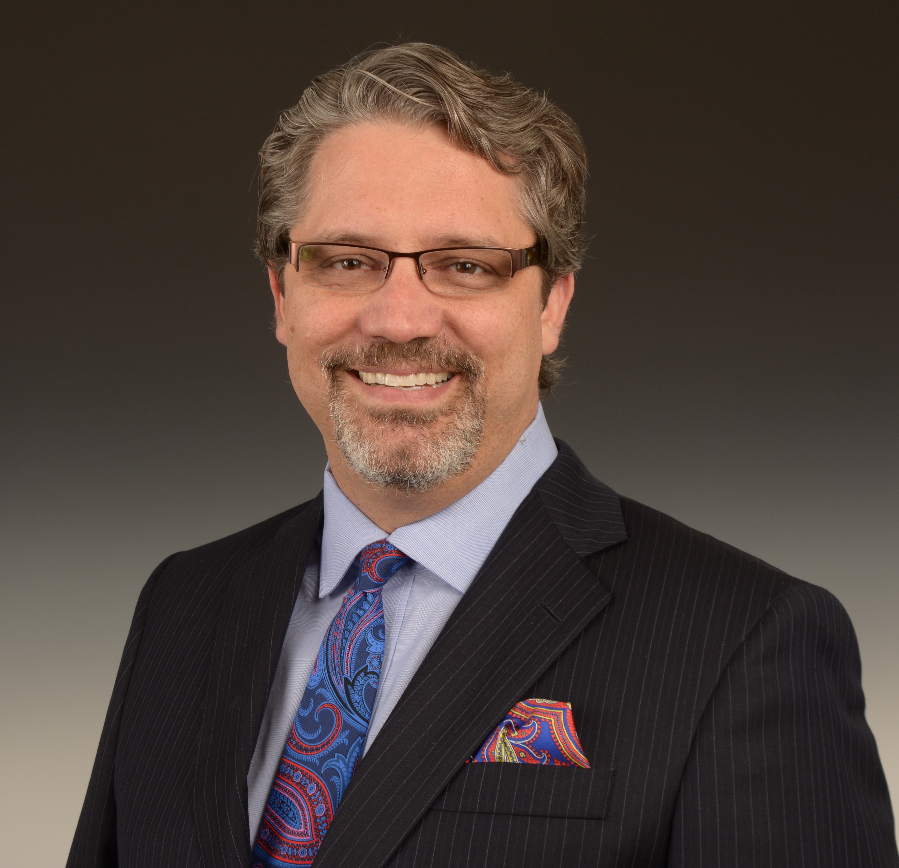
The Academy of Microscope Enhanced Dentistry (AMED) has worked to help inform and educate dentists and the public on the benefits of using the microscope in all areas of dental practice. With Members in 37 countries, this international organization is now helping dental schools introduce the microscope to their undergraduate dental students, non-endodontic residents and faculty. Dr. Linger was elected President of the Academy for 2018.
Microscope Centered Practice
Dr. Linger's office is one of the few microscope centered practices in world, performing routine dental work including dental hygiene with the microscopes.
Dr. Linger has been a pioneer in the use of more sophisticated imaging tools like 3D imaging in the placement of dental implants.
Please contact us to learn more.


